Inside every living organism, a complex and silent conversation is constantly taking place. Cells communicate with one another and respond to their environment through a series of intricate signalling pathways. At the heart of many of these processes is the calcium ion. Acting as a universal second messenger, calcium regulates everything from muscle contraction and neurotransmitter release to gene expression and cell death. To understand how a drug or a biological stimulus affects a cell, scientists need a way to eavesdrop on these calcium signals. This is where the calcium flux assay becomes an indispensable tool in the modern laboratory.
A calcium flux assay allows researchers to monitor the movement of calcium ions into or out of the cytoplasm in real time. By observing these fluctuations, or ‘fluxes’, scientists can determine how cells respond to specific triggers. Whether it is a new pharmaceutical compound intended to treat heart disease or a chemical signal that prompts an immune response, the ability to measure these changes with high precision provides a window into the very mechanics of life. It is not just about seeing that a change happened; it is about understanding the timing, the magnitude, and the duration of that change to build a complete picture of cellular behaviour.
How a calcium flux assay works in the lab
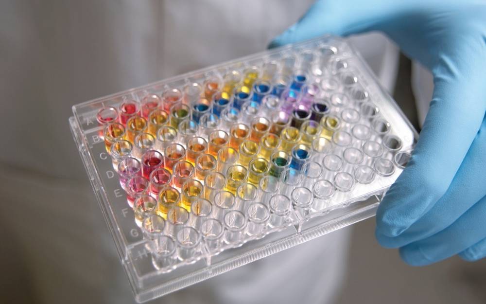
The fundamental principle behind this assay is the use of specialised fluorescent dyes or genetically encoded indicators that change their light-emitting properties when they bind to calcium ions. The process is elegant yet requires careful optimisation to ensure the data produced is both accurate and reproducible. Typically, the workflow involves several key stages that must be meticulously managed.
- Cell Preparation: Cells are cultured and organised into microplates, ensuring they are at the correct density and physiological state for the experiment.
- Dye Loading: A calcium-sensitive fluorescent dye is introduced to the cells. These dyes are often modified with acetoxymethyl (AM) esters, which allow them to cross the cell membrane easily. Once inside, intracellular enzymes cleave the ester groups, trapping the dye within the cell.
- Incubation: The cells are incubated to allow the dye to distribute evenly and for the cellular environment to stabilise.
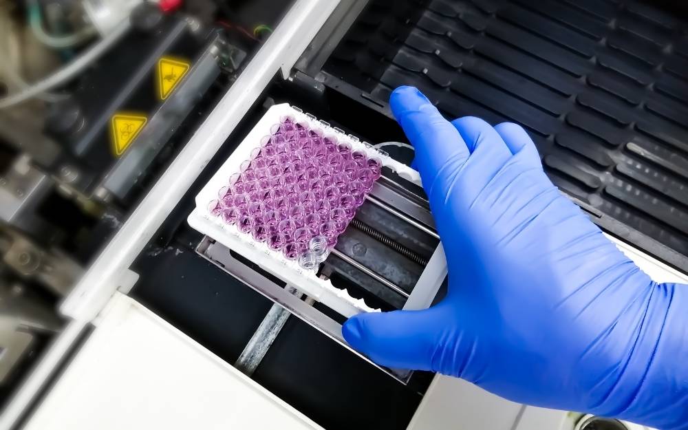
- Stimulation and Detection: A stimulus, such as a drug candidate or a ligand, is added to the cells. A plate reader or an imaging system then measures the change in fluorescence as calcium levels rise or fall.
The beauty of this method lies in its kinetic nature. Unlike many assays that provide a single ‘snapshot’ of a cell’s state at the end of an experiment, the calcium flux assay provides a continuous stream of data. This allows researchers to see the immediate ‘spike’ in calcium and the subsequent recovery, which is vital for understanding the pharmacodynamics of a compound.
Choosing the right fluorescent probes
Not all calcium indicators are created equal, and selecting the right one is critical for the success of the study. Probes are generally categorised into two main types: single-wavelength indicators and ratiometric indicators. Each has its own set of advantages depending on the specific requirements of the research and the equipment available in the facility.
Single-wavelength indicators, such as Fluo-4 or Cal-520, are popular because they offer very high sensitivity and a large increase in fluorescence intensity upon binding to calcium. They are excellent for high-throughput screening where speed and signal strength are the primary concerns. However, they can be sensitive to variations in dye loading or cell thickness, which can sometimes lead to inconsistencies in the data if not properly controlled.
Ratiometric indicators, like Fura-2 or Indo-1, work differently. They shift their excitation or emission spectra when they bind to calcium. By measuring the ratio of light at two different wavelengths, researchers can cancel out variables such as uneven dye loading, photobleaching, or changes in cell volume. This makes them highly accurate for quantitative measurements of absolute calcium concentrations, though they often require more sophisticated optical setups and can be slower to process than single-wavelength alternatives.
Why researchers still depend on this method
Despite the emergence of many new technologies in molecular biology, the calcium flux assay remains a cornerstone of drug discovery, particularly for studying G-protein coupled receptors (GPCRs) and ion channels. GPCRs are the targets for an enormous percentage of modern medicines, and many of them signal through the mobilisation of intracellular calcium. By using these assays, pharmaceutical companies can screen thousands of compounds rapidly to identify ‘hits’ that either activate or inhibit these receptors.
Beyond simple screening, these assays are vital for safety pharmacology. For instance, in cardiac research, observing calcium handling is essential for predicting whether a drug might cause dangerous arrhythmias. If a compound disrupts the normal rhythm of calcium release and reuptake in heart cells, it is a significant red flag for potential cardiotoxicity. This early insight saves time and resources, ensuring that only the safest compounds move forward into clinical trials.
The move toward high-throughput screening
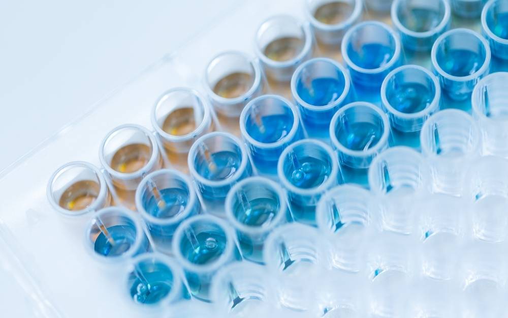
In the competitive world of drug development, speed is essential. The industry has moved toward high-throughput screening (HTS) formats, where calcium flux assays are performed in 384-well or even 1536-well plates. This level of automation allows for the testing of vast libraries of chemicals in a fraction of the time it would take using traditional methods. Several factors contribute to the success of HTS in this context:
- Automated Liquid Handling: Precise robotics ensure that reagents and compounds are added at the exact same moment across all wells, which is crucial for capturing fast kinetic responses.
- Advanced Plate Readers: Modern instruments can read entire plates simultaneously, capturing the rapid rise in fluorescence that occurs within seconds of stimulation.
- Data Integration: Sophisticated software can automatically calculate peak heights, area under the curve, and decay rates, turning raw light signals into actionable biological insights.
Navigating the technical hurdles
While the concept is straightforward, executing a high-quality assay requires navigating several technical challenges. One common issue is background fluorescence, which can come from the compounds being tested or the media used to grow the cells. To combat this, researchers often use ‘quenchers’—substances that stay outside the cell and suppress any fluorescence not coming from the intracellular dye. This significantly improves the signal-to-noise ratio, making it easier to detect subtle changes in calcium levels.
Another consideration is the choice of cell type. While immortalised cell lines are easy to grow and provide consistent results, they may not always reflect the true biology of a human patient. Increasingly, researchers are turning to primary cells or induced pluripotent stem cell (iPSC)-derived cells. These models offer a more physiologically relevant environment, though they can be more temperamental to work with and require more intensive optimisation of the assay conditions.
Timing is also a critical factor. The peak of a calcium response often occurs within seconds of adding a stimulus. If the detection system is too slow, or if the liquid addition is not perfectly synchronised, the most important part of the data could be missed entirely. This is why the integration of ‘add-and-read’ systems has been such a game-changer for the field, allowing for the seamless transition from stimulation to measurement without any manual intervention.
Looking ahead at cellular monitoring
The future of measuring cellular signals is moving toward even greater complexity and biological relevance. We are seeing a shift toward multi-parametric assays, where calcium flux is measured alongside other indicators, such as membrane potential or metabolic activity, in the same cell at the same time. This ‘multiplexing’ provides a holistic view of cellular health and response that a single measurement simply cannot match. Furthermore, the development of 3D cell culture models and ‘organ-on-a-chip’ technology is pushing the boundaries of how we use these assays to simulate the human body in a laboratory setting. As our ability to model human disease improves, the humble calcium flux assay continues to evolve, remaining a vital part of the toolkit for anyone looking to understand the complex, shimmering signals that drive life at the cellular level.

Charlotte Reid is a seasoned travel writer with a passion for exploring unique accommodations and uncovering hidden gems in the hospitality industry. With years of experience in lifestyle journalism, she focuses on creating engaging content that helps readers find the perfect getaway spots. Her work emphasizes comfort, charm, and the personal touches that make every stay memorable.